SNSF Scientific Image Competition
Get your cameras! Give Swiss research a face.
The SNSF Scientific Image Competition encourages researchers working in Switzerland to present their works to the public and the media. Photographs, images and videos will be rated in terms of their aesthetic quality and their ability to inspire and amaze, to convey or illustrate knowledge, to tell a human story or to let us discover a new universe.
You can find all competition entries, past and present, here:
Winners 2026
The jury has awarded four first prizes and sixteen distinctions out of the 314 submitted entries.
Category 1 – Object of study
 Claire Galloni © Claire Galloni
Photo-ethnography hybridising contemporary arts, by Claire Galloni d'Istria
Postdoctoral researcher, University of Geneva
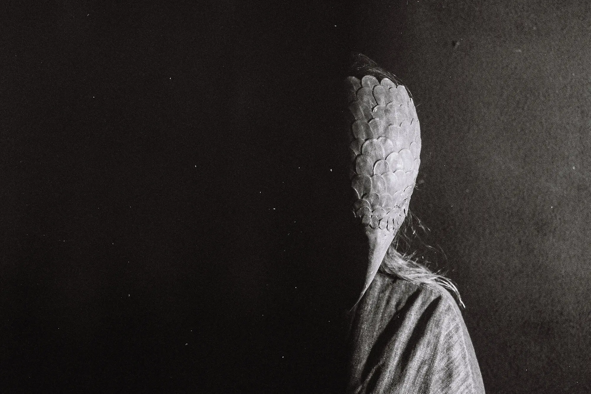
The experimental photo-ethnography that I employ combines art and ethnography to explore different ways of inhabiting Alpine environments. My colleague Anna Sakowicz wears a bird mask borrowed from the collection of the Théâtre Am Stram Gram in Geneva, created by the artist Werner Strub, introducing a non-human presence through a theatrical device.
I use photography and sound in a multimodal anthropological approach to explore the way in which human and non-human movements and practices are currently being reconfigured in alpine environments. I study how these territories are experienced, negotiated and redefined in a context of rapid ecological change.
This image of a hybrid creature questions how human identities can also be understood in relation to animal entities – in this case, birds. It reveals the fragility of our relationships through a staged composition that combines ethnographic research and contemporary visual art practices.
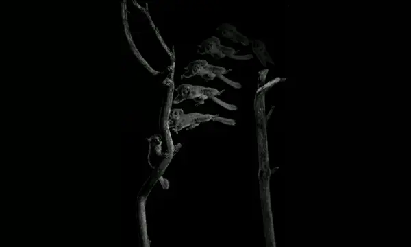
Jury’s commentary │ With its pared-down, dramatic depiction of a hybrid – almost mythological – creature, this striking photograph showcases the originality of the research approach. The radical framing highlights a slightly disquieting darkness that seems likely to be concealing other hidden figures.
Category 2 – Women and men of science
 Mirjam Widmer © Mirjam Widmer
Braving the water, by Mirjam Widmer
Speleologist, Swiss Speleological Society and student, Zentrum Bildung
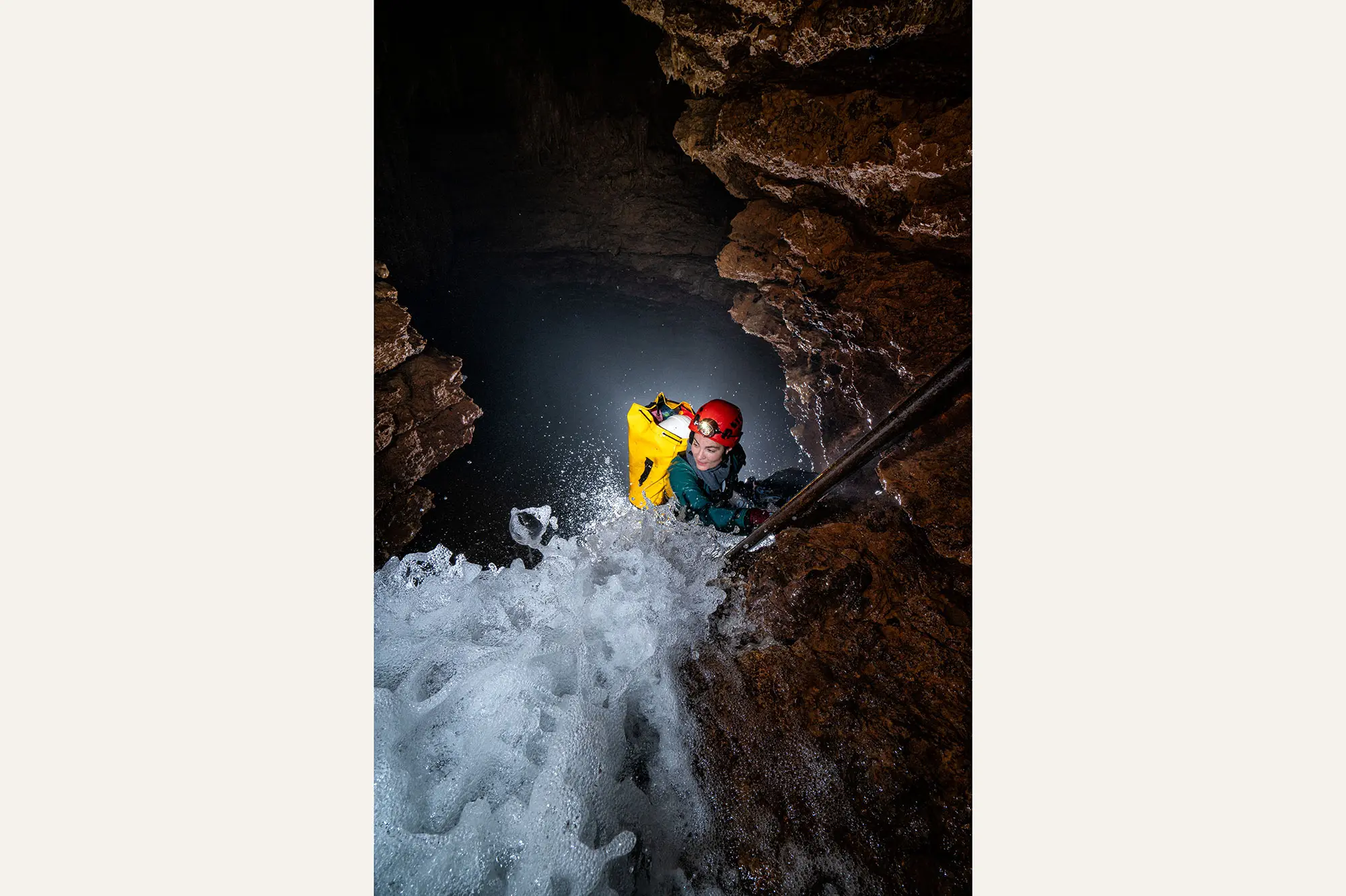
The photo shows cave scientist Ainhoa Val at the Grande Cascade in the karst cave known as the Grotte de Milandre, near Boncourt (Jura). This passage is extremely loud and very wet, and the image seeks to convey the power of the flowing water.
All the research equipment had to be transported to the study site of the Swiss Institute of Speleology and Karstology's CaveSeds project, about an hour and a half from the cave's entrance. To ensure safety, access is only possible under specific environmental conditions, including an acceptable concentration of CO₂ inside the cave and a sufficiently low water level. The photo was taken using two external flashes triggered remotely.
Jury's commentary │ This spectacular photograph plunges us into a world that seems almost inaccessible. It lets us feel the power and noise of the current – while hinting at the positive emotion of the protagonist – in a technical tour de force that masters lighting and composition.
Category 3 - Locations and instruments
 Jayant Abhir © Jayant Abhir
Moonshot, by Jayant Abhir
PhD candidate, ETH Zurich
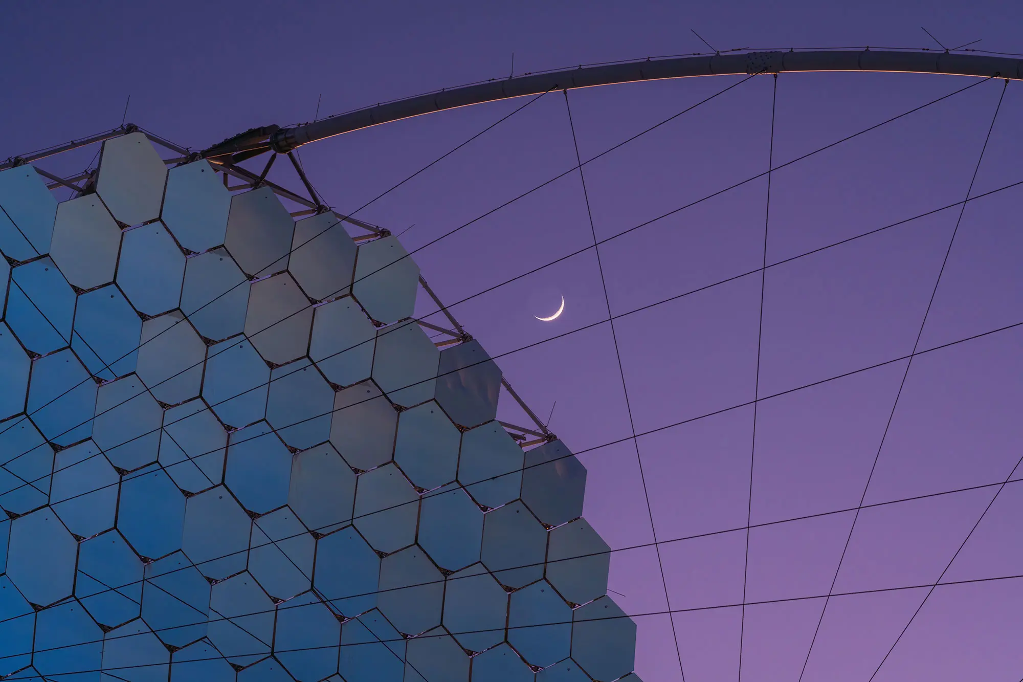
The moon appears framed by the mechanical structure and support wires of the CTAO LST1 telescope at an altitude of 2,200 metres at the Roque de los Muchachos Observatory in La Palma. The moon’s topography is highlighted by the terminator line – the boundary eparating the bright and shaded parts of its surface – even revealing the edges of some of its craters.
The LST1 is the first in an array of Cherenkov telescopes that are 23 metres in diameter. Its 1.5-metre mirror segments reflect the sky at dusk, creating an inverted image. In some of the mirrors at the very top of the structure, you can see the cloud inversion layer that forms at a lower altitude than the site, a meteorological phenomenon that makes this location ideal for astronomy.
Jury's commentary │ A sophisticated composition that juxtaposes the mechanical and the natural, the near and the far, the abstract and the figurative.
Category 4 - Videos
 Inés Segovia Campos © Inés Segovia Campos
Links
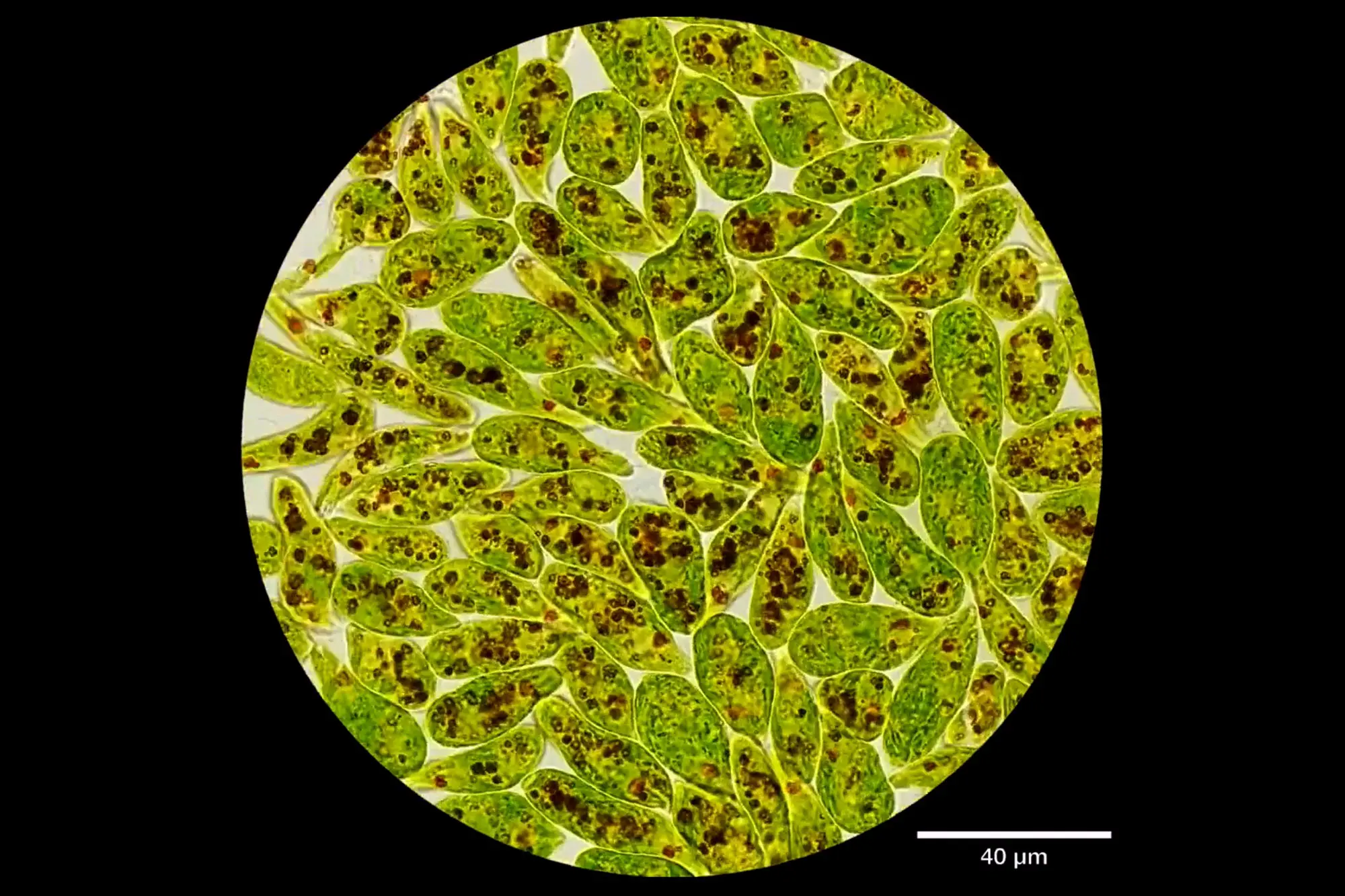
A living crowd at the microscopic scale, by Inés Segovia Campos
Postdoctoral researcher, University of Geneva
This video (accelerated 20x) shows the collective motion of densely packed Euglena gracilis cells, observed under a microscope using a 60x oil-immersion objective. These single-celled organisms blur the line between plants and animals: they contain chloroplasts and can photosynthesise like plants, and like animals, they feed heterotrophically, i.e. by consuming external organic matter.
Euglena does not have a rigid cell wall and continuously changes shape to navigate through dense or viscous environments. Its ability to rapidly adjust its metabolism and behaviour makes it a valuable model organism in ecotoxicology, which is widely used as an early indicator of pollution and environmental stress in aquatic ecosystems. This video reveals the hidden dynamics of microscopic life, where apparent chaos masks remarkable adaptability.
Jury's commentary │ Cells jostle, contort and move about as best they can in a classic, precise shot in intense colours that reveals a microscopic world no less crowded than our modern cities.
Links
Winners 2017–2025
Online gallery
All of the images are presented in our online gallery. Follow the images from the competition on Instagram @swissnationalsciencefoundation and on X using the hashtag #SwissScienceImage.
Participation
Participation requirements
All scientists working at a research institution in Switzerland are eligible to participate. The works must have been produced less than 12 months before the deadline for submitting entries.
Submission
Researchers who wish to take part in the competition must fill in the online form.
Categories of the competition
Each participant may submit from 1 to 5 entries, in one or more of the following categories:
1) Object of study (image)
From the microcosm to the macrocosm, images of the research object captured by scientists using a camera or generated by a computer.
2) Women and men of science (photographs)
Photographs of research in practice, presented by and featuring those conducting it.
3) Locations and instruments
Photographs of the surroundings in which scientists take measurements, generate data and make discoveries, and of the instruments they use while doing so.
4) Video loop
Chronophotography, video or animated gif, documenting some aspects of categories 1 to 3.
Technical details
Photography
Digital image file obtained from a camera. Format: JPEG or TIFF. Maximum size: 100 MB. Minimum resolution: 2000 x 3000 pixels (16.9 x 25.4 cm to 300 dpi). Digital touching up permitted.
Image
Digital image file taken from a camera or computer-generated from data obtained through observation or computer simulation (excluding explanatory infographics). Others: see "Photographs", above.
Video
Digital video file taken from a camera or computer-generated from data obtained through observation or computer simulation (excluding explanatory infographics). Formats: GIF, AVI, MP4 (edited in a loop). Maximum size: 300 MB. Duration: from 3 to 15 seconds. Minimum resolution: 480 x 720 pixels (DVD resolution). Digital touching up permitted.
Terms of use
The participants retain their copyright. They authorise the publication of the submitted images under a CC-BY-NC-ND licence (https://creativecommons.org/licenses/by-nc-nd/3.0/): unaltered images can be used freely for non-commercial purposes as long as they are credited as the creator of the image.
About the competition
The competition is held annually. An international jury will meet at the beginning of the year and award a CHF 1,000 prize in each category for the winning entry, as well as CHF 250 for each distinction. The award-winning works are announced at the end of April, displayed in an exhibition at the Biel/Bienne Festival of Photography and made available to the public and the media, as well as to scientific institutions.
The competition has multiple aims: to highlight the growing role of images in scientific research, to reveal how scientific work is conducted and to give a face to the researchers conducting it. The competition also aims to encourage the media to use more images in their science coverage and make them accessible to the public through exhibitions.
We encourage researchers to pick up their camera and document the – often unusual – environment in which they work, and to give their colleagues a face.
Jury 2026
The jury includes international experts in the fields of photography, museums, media and research from around the world.
Chair
Patrick Gyger, director of Plateforme 10 (Switzerland)Members
Jessica Hallett, media editor, Nature (England)
Andri Pol, photographer (Switzerland)
Tess de Ruiter, art-science curator, Rotterdam (Netherlands)
Aurélie Saliba, manager crowdsourcing for Adobe Stock (Germany)Award ceremony, exhibition and online galleries
The award ceremony will take place in May 2026 during the Biel/Bienne Festival of Photography, where a selection of the works will be exhibited.
The images are presented at other exhibitions as well as online:
People’s prize 2017–2021
In March 2021, the public voted for its favourites from among 50 photographs and 15 videos. The preselection had been made in February 2021 by 20 photography students of Arts College Bern/Biel.
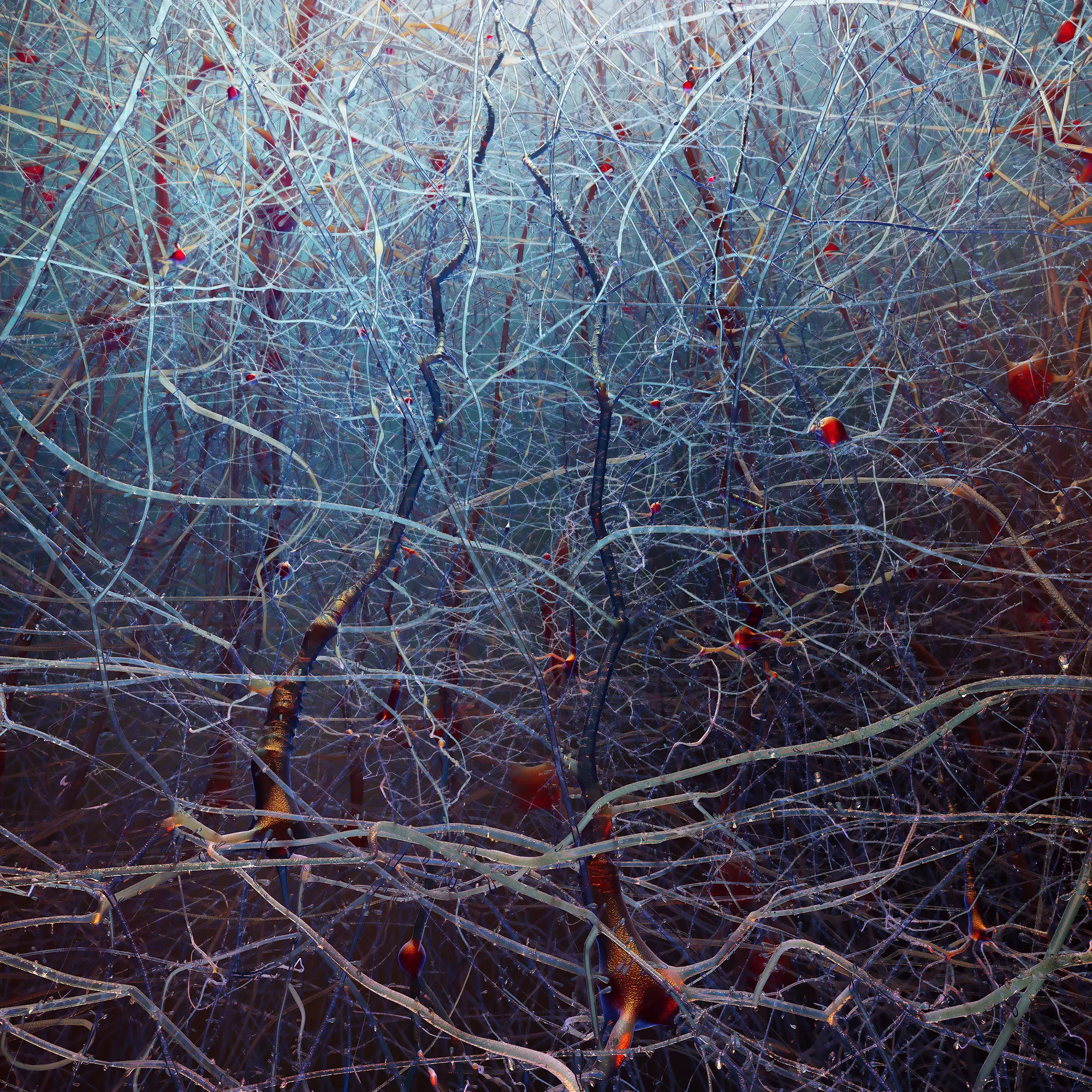
A view from inside the neocortical forest (2017)
Nicolas Antille (EPFL)
Jump! (2021) - Video
Daniel Huber (University of Geneva)
News
Year